")
Understanding 3D Immunohistochemistry (IHC)
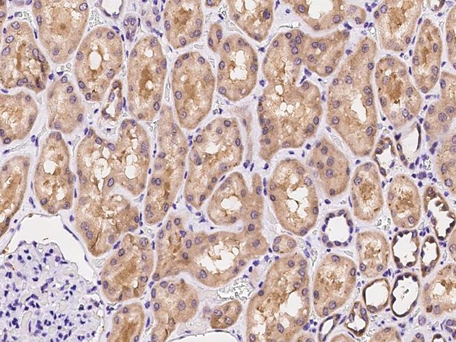
Immunohistochemistry (IHC) is a powerful technique used in pathology to visualize the presence and localization of specific proteins within tissue sections. Traditionally, IHC has been performed on 2D sections, but the advent of 3D immunohistochemistry (3D IHC) has revolutionized the field by providing a more comprehensive view of tissue architecture and protein distribution.
What is 3D IHC?
3D IHC is a technique that allows for the visualization of tissue samples in three dimensions, providing a more accurate representation of the spatial relationships between cells and proteins. This is achieved by using specialized staining methods and imaging techniques that capture the entire volume of the tissue sample.
")
Staining Techniques
One of the key components of 3D IHC is the staining method. There are several staining techniques available, each with its own advantages and limitations. Some of the most commonly used staining methods include:
| Staining Method | Description |
|---|---|
| Peroxidase-based staining | Utilizes peroxidase enzymes to detect specific proteins. This method is widely used due to its high sensitivity and specificity. |
| Alkaline phosphatase-based staining | Similar to peroxidase-based staining, but uses alkaline phosphatase enzymes. This method is often used for proteins that are not easily detected by peroxidase-based staining. |
| Fluorescence-based staining | Utilizes fluorescent dyes to visualize proteins. This method is particularly useful for multiplexing, where multiple proteins can be visualized simultaneously. |
Imaging Techniques
Once the tissue sample is stained, it needs to be imaged to visualize the 3D structure. There are several imaging techniques available, each with its own advantages and limitations. Some of the most commonly used imaging techniques include:
| Imaging Technique | Description |
|---|---|
| Confocal microscopy | Uses a laser to scan the tissue sample and capture images at different depths. This allows for the creation of 3D reconstructions of the tissue. |
| Micro-computed tomography (micro-CT) | Utilizes X-rays to create cross-sectional images of the tissue sample. These images can then be used to create 3D reconstructions. |
| Optical projection tomography (OPT) | Utilizes a rotating stage and a camera to capture images of the tissue sample from multiple angles. These images are then used to create a 3D reconstruction. |
Applications of 3D IHC
3D IHC has a wide range of applications in both research and clinical settings. Some of the most notable applications include:
-
Studying the spatial distribution of proteins within tissue samples
-
Identifying the cellular context of protein expression
-
Assessing the progression of diseases, such as cancer
-
Developing new diagnostic and therapeutic strategies
Advantages of 3D IHC
Compared to traditional 2D IHC, 3D IHC offers several advantages:
-
Improved spatial resolution, allowing for a more accurate representation of tissue architecture
-
Increased sensitivity and specificity, leading to better detection of proteins
-
Ability to visualize the entire volume of the tissue sample, providing a more comprehensive view of protein distribution
Challenges and Future Directions
Despite its many advantages, 3D IHC still faces several challenges. Some of the main challenges include:
-
Development of new staining and imaging techniques to


